DIAGNOSTICS
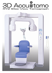
Digital Volume Tomography with the 3D Accuitomo
My practice features one of the 18 units installed in Germany.
Conventional x-ray imaging produces two-dimensional projections of anatomical structures. The benefit of volume tomography is that it additionally portrays the third dimension; images may be viewed in extremely thin layers so that anatomical structures do not conceal one another.
Computed tomography (CT) was developed because it was difficult to draw conclusions from traditional x-ray diagnostics.
What are the advantages of the 3D Accuitomo over common x-ray imaging?
- Representation of the area being examined in all three spatial dimensions
- Extremely high resolution and excellent image quality
- Thinly layered images, resulting in a clear view of anatomical structures
- Lower dose of radiation
- Shorter exposure time (only 17 seconds)
- Comfortable chair and a more relaxed atmosphere
You will need the current Adobe Flash Player to watch the videos. If you do not already have it on your computer, you can download it here: www.adobe.com
top
X-Ray Procedure
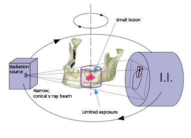
The patient’s chair is moved along three axes so that the area to be examined is in the correct position. The detector arm then rotates 360° around the patient’s head.
top
When is the 3D Accuitomo helpful?
- In all cases where conventional x-ray images do not result in a clear diagnosis
- In analyzing hidden roots for difficult root canal procedures
- To precisely determine whether damaged teeth can be saved
- To evaluate the maxillary sinus and the mandibular nerve, as well as the distance between these anatomical structures
- In diagnosis prior to implant procedures and to measure the dimensions of the implant bed
Berlin 2010, Dr. med. Dr. med. dent. Herbert Kindermann